A Dural Arteriovenous Fistula (DAVF) is an abnormal connection between arteries and veins located within the dura mater, the outer protective covering of the brain. Although relatively rare, DAVFs can lead to serious neurological complications, including stroke, if left untreated. Early diagnosis and timely treatment at a specialized stroke centre can significantly reduce risks and improve long-term neurological outcomes.
What Is a Dural Arteriovenous Fistula?
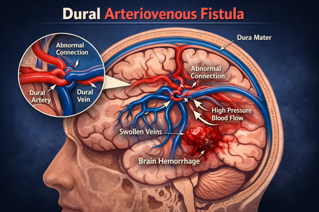
A Dural Arteriovenous Fistula occurs when abnormal channels form between dural arteries and dural venous sinuses or cortical veins. Normally, blood flows from arteries to capillaries and then to veins. In DAVF, this normal circulation is bypassed, causing high-pressure arterial blood to flow directly into veins.
This abnormal blood flow can increase venous pressure in the brain and lead to serious complications such as intracranial hemorrhage, neurological deficits, or stroke.
Because of these potential risks, evaluation by an experienced brain stroke physician is essential to determine the severity and appropriate treatment approach.
Causes and Risk Factors
The exact cause of DAVF is not always known, but several conditions may contribute to its development:
- Venous sinus thrombosis
- Previous brain surgery or trauma
- Head injury
- Infections affecting the brain coverings
- Congenital vascular abnormalities
In many cases, DAVFs develop after blockage or thrombosis of a venous sinus, which triggers abnormal vascular connections.
Symptoms of Dural Arteriovenous Fistula
Symptoms vary depending on the location and severity of the fistula. Some patients may experience mild symptoms, while others may develop severe neurological complications.
Common symptoms include:
- Pulsatile tinnitus (whooshing sound in the ear)
- Persistent headaches
- Visual disturbances
- Seizures
- Neurological weakness or numbness
- Cognitive changes
Diagnosis of DAVF
Accurate diagnosis requires advanced imaging techniques to identify the abnormal vascular connections and evaluate blood flow patterns.
Common diagnostic tools include:
- MRI and MR Angiography
- CT Angiography
- Digital Subtraction Angiography (DSA)
DSA remains the gold standard for diagnosing DAVF because it provides detailed visualization of the arteries, veins, and abnormal fistula connections.
Treatment Options for DAVF
The goal of treatment is to eliminate the abnormal artery-vein connection and restore normal blood circulation in the brain.
Endovascular Embolization
The most common and effective treatment for DAVF is endovascular embolization, a minimally invasive procedure performed through blood vessels.
During this procedure:
- A small catheter is inserted through the wrist or groin artery.
- The catheter is navigated to the abnormal fistula in the brain.
- Special embolic materials such as liquid embolic agents or coils are used to close the abnormal connection.
This technique allows precise treatment while avoiding open brain surgery.
Surgical Treatment
In certain cases where endovascular therapy is not feasible, surgical disconnection of the fistula may be required.
Radiosurgery
For selected low-risk DAVFs, stereotactic radiosurgery may be used to gradually close the abnormal vessels over time.
Why Early Treatment Is Important
Untreated DAVFs can lead to progressive neurological damage. High-risk fistulas, especially those involving cortical venous reflux, carry a significant risk of intracranial hemorrhage and stroke.
Early intervention at a specialized Brain Stroke Centre helps prevent life-threatening complications and ensures better recovery outcomes.
Expertise Matters in DAVF Treatment
Managing complex neurovascular conditions like DAVF requires specialized expertise and advanced imaging technology. Evaluation and treatment by an experienced neurologist ensure accurate diagnosis, proper risk assessment, and the most effective treatment approach.
Comprehensive care at a modern brain stroke centre includes multidisciplinary evaluation, minimally invasive interventions, and long-term follow-up to protect brain health and reduce the risk of stroke.
Conclusion
Dural Arteriovenous Fistulas are rare but potentially dangerous vascular conditions that can lead to serious neurological complications if untreated. Early diagnosis, proper risk evaluation, and advanced treatment techniques are essential for preventing brain damage and stroke.
Seeking care at a specialized brain stroke centre under the guidance of a skilled brain stroke physician ensures timely intervention, safer treatment, and improved long-term neurological outcomes.