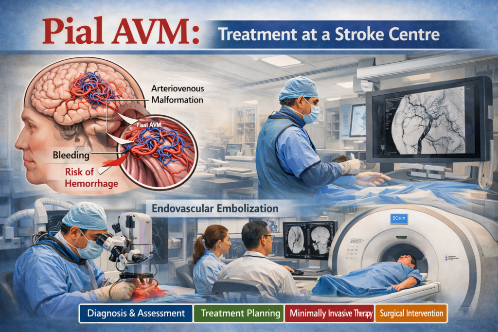
A Pial AVM (Pial Arteriovenous Malformation) is a complex and potentially life-threatening vascular disorder of the brain. It occurs when arteries and veins connect abnormally without the normal capillary network, leading to high-pressure blood flow that can damage delicate brain tissue.
This condition significantly increases the risk of bleeding in the brain and brain stroke, especially if left untreated. Early detection and timely management at a specialized stroke centre or brain stroke centre are crucial for preventing serious complications.
With the expertise of a qualified brain stroke physician, patients can receive advanced diagnostic evaluation and personalized treatment plans that improve survival and recovery outcomes.
What Is a Pial AVM?
A Pial Arteriovenous Malformation is a type of brain AVM located on the surface (pial layer) of the brain. It consists of a tangled cluster of abnormal blood vessels called a nidus, where arteries directly connect to veins.
Normal vs Abnormal Blood Flow
Normal Circulation:
- Arteries carry oxygen-rich blood
- Capillaries regulate and slow blood flow
- Veins return blood to the heart
In Pial AVM:
- Capillaries are bypassed
- Blood flows rapidly from arteries to veins
- Veins are exposed to high pressure
- Risk of rupture and bleeding increases
Causes of Pial AVM
1. Congenital Causes (Most Common)
- Develops before birth due to abnormal blood vessel formation
- Exact cause remains unclear
- Often diagnosed later in life when symptoms appear
2. Rare Acquired Causes
- Severe head trauma
- Vascular abnormalities or disorders
- Previous brain infections (rare cases)
Risk Factors
Although Pial AVMs are usually congenital, certain factors may increase complications:
- High blood pressure
- Smoking
- Family history of vascular disorders
- Stress or physical strain
- Age (symptoms often appear between 20–40 years)
Symptoms of Pial AVM
Symptoms depend on the size, location, and whether the AVM has ruptured.
Common Symptoms
- Persistent or severe headaches
- Seizures (one of the most common signs)
- Weakness or numbness in the body
- Difficulty speaking or understanding speech
- Vision disturbances
- Dizziness or loss of balance
Symptoms of Ruptured Pial AVM (Medical Emergency)
- Sudden severe headache (“worst headache of life”)
- Nausea and vomiting
- Loss of consciousness
- Sudden paralysis or weakness
- Seizures
- Confusion or altered mental state
These symptoms may indicate a brain hemorrhage or brain stroke and require immediate care at a brain stroke centre.
Complications of Pial AVM
If left untreated, Pial AVMs can lead to serious complications:
- Brain hemorrhage (bleeding)
- Increased intracranial pressure
- Permanent neurological damage
- Cognitive impairment
- Disability or paralysis
- Life-threatening Brain Stroke
Diagnosis of Pial AVM
Accurate and early diagnosis is essential for effective treatment planning.
Advanced Diagnostic Methods
MRI (Magnetic Resonance Imaging)
- Provides detailed brain images
- Identifies abnormal blood vessels
CT Scan
- Detects bleeding in the brain
- Useful in emergency situations
Cerebral Angiography (Gold Standard)
- Detailed imaging of brain blood vessels
- Helps locate the AVM precisely
- Essential for treatment planning
Additional Evaluation
- Neurological examination
- Assessment of patient history and symptoms
Treatment Options for Pial AVM
Treatment depends on multiple factors such as size, location, and rupture risk.
1. Endovascular Embolization
- Minimally invasive procedure
- Catheter inserted through blood vessels
- Special material used to block abnormal vessels
- Often used before surgery or radiosurgery
2. Microsurgical Removal
- Surgical removal of the AVM
- Provides immediate elimination of the abnormality
- Best for accessible AVMs
3. Stereotactic Radiosurgery
- Non-invasive radiation therapy
- Gradually closes abnormal blood vessels
- Suitable for smaller or deep AVMs
4. Multimodal Treatment Approach
- Combination of embolization, surgery, and radiation
- Offers better outcomes for complex AVMs
Importance of Early Treatment at a Stroke Centre
Getting treated at a specialized stroke centre or brain stroke centre offers significant advantages:
- Early and accurate diagnosis
- Access to advanced imaging technologies
- Minimally invasive treatment options
- 24/7 emergency stroke care
- Experienced brain stroke physicians
- Multidisciplinary treatment approach
Timely care reduces the risk of complications and improves long-term recovery.
Role of a Brain Stroke Physician
A brain stroke physician plays a critical role in managing Pial AVM:
- Diagnosing complex brain vascular conditions
- Planning personalized treatment strategies
- Preventing stroke and complications
- Monitoring recovery and rehabilitation
- Providing long-term follow-up care
Their expertise ensures safe, effective, and comprehensive patient care.
When to Seek Immediate Medical Attention
Visit a brain stroke centre immediately if you experience:
- Sudden severe headache
- Seizures
- Weakness or numbness on one side
- Difficulty speaking or understanding
- Vision problems
- Loss of consciousness
Early intervention can save lives and prevent permanent brain damage.
Lifestyle and Prevention Tips
While Pial AVMs cannot always be prevented, patients can reduce risks by:
- Controlling blood pressure
- Avoiding smoking and alcohol
- Managing stress levels
- Following prescribed medications
- Attending regular medical check-ups
Conclusion
A Pial AVM is a serious and potentially life-threatening brain condition that requires prompt medical attention. Without proper treatment, it can lead to severe complications such as brain hemorrhage and brain stroke.
However, with early diagnosis and advanced treatment at a specialized stroke centre or brain stroke centre, patients can achieve significantly better outcomes.
Under the guidance of an experienced brain stroke physician, individuals receive accurate diagnosis, personalized care, and long-term management strategies that protect brain health and reduce stroke risk.